
Dimensional measurements of nanoparticles are extremely important for their applications in a diverse range of fields. The unique properties of nanoparticles depend strongly on their size, so it’s extremely important to be able to characterize, and control nanoparticle sizes. For example, an extremely small change in the dimension of a quantum dot nanoparticle will give rise to dramatically different photoluminescence characteristics. in the case of metallic nanoparticles, the size of the particle will alter light absorption and scattering properties, which are highly important for many applications, including in disease diagnosis and therapy (1). Many other properties depend on nanoparticle size, including magnetic and mechanical properties, interactions with cells and tissues, etc. The shape of nanoparticles also has a strong effect on these properties.
Thus, in nanoscience, it is crucial to have a tool that can simply characterise particles size and shape of nanoparticles, irrespective of the material of which they are composed, with sub-nanometer resolution. The figure below shows AFM imaging of a variety of nanoparticle types.
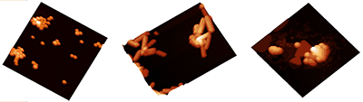
Figure above shows (L-R) silica nanospheres, organometallic nanorods, and gold nanotriangles all imaged by AFM. Adapted from figure 7.11 of Eaton and West, Atomic Force Microscopy(2)
AFM fulfils these requirements, z-axis (height) measurements in AFM can be accurate to within 0.1 nm. AFM also has several other advantages for characterisation of nanoparticles:
-
AFM can characterise properties other than simple topography such as magnetic, mechanical properties.
-
AFM can image both organic and inorganic components, e.g. interaction between nanoparticles and biomolecules (3)
-
Unlike light scattering techniques, AFM (like other microscopic techniques is suitable to characterise mixtures of nanoparticles (see example below). For techniques such as DLS; mixed samples will only give an average results at best; often only the larger component will be detected.
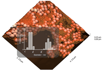
Figure above shows an AFM image of a mixed population of nanoparticles, with (inset) a histograms of the diameters of the nanoparticles, measured form the images obtained.
-
Another advantage compared to light scattering techniques is the ability to characterise shape as well as size.
-
AFM gives strong contrast on any material; electron microscopy often gives poor contrast with organic materials
In conclusion, atomic force microscopy, AFM is a highly suitable technique to characterise the size and shape of wide variety of nanoparticles.
This article was partially adapted from Eaton and West, Atomic Force Microscopy.
References
1. Baptista et al., Nanoparticles in Molecular Diagnostics, Progr. Mol. Biol. Trans. Sci. 104, 427-488 (2011).
2. Eaton and West, Atomic Force Microscopy, Oxford University Press, 2010. ISBN: 978-0199570454.
3. Eaton et al, Imaging Gold Nanoparticles for DNA Sequence Recognition in Biomedical Applications, IEEE Trans. NanoBioScience 6(4), 282 - 288 (2007).
All text and images copyright 2010 and 2015 Peter Eaton. No use is permitted without explicit permission.