In my job as a researcher using AFM, I often act as a scientific reviewer for papers describing AFM experiments. I also collaborate with many groups and spend a lot of time revising and correcting articles which contain AFM data. In this two part article, I’ll describe how you should go about preparing a scientific paper which contains AFM data.
NOTE: Some of this content is not really specific to AFM articles, but could be applied to any experimental scientific report. These parts should already be known to you if you are writing an scientific article, but many researchers are never taught how to correctly prepare a scientific article before they start writing them. For more general guides on preparing scientific articles, check here.
What are the overall requirements for an AFM paper?
-
Experimental conditions should be well described
-
Data should be presented in a useful format
-
Reproducibility should be addressed
-
Conclusions should be justified
These requirements may seem like “common sense”, but you might be surprised by the number of articles which are published but do not meet some, or even all of these criteria!
-
Experimental conditions:
What should this include:
Imaging mode used. This is rather important, since the results can be different from mode to mode. It also helps to make your data reproducible. Note that not all instruments use the same terms to refer to specific modes :This is particularly the case for tapping mode. It can be useful to use both the manufacturer’s name, and the most commonly used name for example: Imaging was carried out in AC-AFM (Tapping) mode.”
Instrument used. You should name the instrument mode and the manufacturer. You can also include the version of the instrument model, if applicable. Some journals still request the location of the company, but this is becoming optional e.g., you might write “Bruker Multimode 8 (Bruker, Newhaven, CA)”.
Probe used. The type of probe used can determine the type of information available, as well as the interaction force. Since there are many probes available on the market, it’s also useful to say the (approximate) frequency of the probe, which gives some idea of the type of probe, without he use looking up the details. E.g. you might say “AppNano ACT probes with around 300 kHz frequency were used”.
Software used. All AFM images require image processing, or analysis before presentation. It is a good idea to describe the software used for processing of the AFM data. You might also describe the processes used for analysis and /or data processing. E.g. you might say “Gwyddion 2.49 software was used for processing the images, and Roughness was calculated with the Gwyddion 'Statistical Parameters' tool".
Amount of data collected, and statistical significance. This issue is addressed below. However, it's worth remembering, that the microscopy images you collect should be representative of the sample(s). A part of the “materials and methods” section should be explaining this. For example, you might write “At least six areas per sample were imaged, and representative images are shown here.” If you have a variety of different images, you could add additional images to a “supporting information” section.
-
Presenting data in a useful format
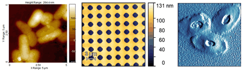
There are many different types of AFM images, and different ways to display them. Very often the instrument will produce several different image channels, such as height, phase, etc. The most commonly used channel is probably height, since this is the only one with three-dimensional data. However, sometimes, it’s hard to make out the sample details in height images. For this reason, other channels, such as deflection or phase are often used. I recommend that the height image, always come accompanied with a z scale (normally it’s a colour gradient), and they should have either an inset scale bar, distance markers on the outside, or the legend must state the scan size (x and y). Examples are shown below.
Does the colour I pick make any difference? No. You can choose which colours you like. For some reason, AFM images are “traditionally” shown in some shade of brown, but any colour scale is fine.
Should Amplitude/Deflection/Phase images have a z scale? No. The z scale on these channels is usually meaningless, as it might be in volts, or degrees, and in most conditions, is not needed. I recommend to remove it to avoid confusion.
Are the details you want to highlight easily visible? If not, the use of arrows to denote specific features in the image is recommended. The arrows should be referenced in the figure legend.
Talking of which, in part 2 of this article, I’ll talk about how a figure legend should be written, and how you should address reproducibility of the data, and draw appropriate conclusions from your results.
All images copyright peter Eaton 2019. Images produced with SPIP and Gwyddion software