
This instrument is supervised by: Peter Eaton, contact: This email address is being protected from spambots. You need JavaScript enabled to view it.


This instrument is a modified TT-AFM from AFM Workshop.
It is equipped with two scanners, enabling large (low resolution ) or small (high resolution) scanners. It has an experimental liquid cell for in-situ measurements.
Here are more detailed specifications:
- Instrument Configuration: Light lever (optical lever) - based sample-scanning AFM
- Sample Sizes:ca. 13x13x5 mm
- Imaging Modes: vibrating (tapping), non-vibrating (contact), phase imaging, lateral force microscopy (friction force microscopy)*
- Imaging Environment: Air or Liquid* (experimental).
- Z-translation: Vertical direct drive (1micron resolution)
- XY Translation: manual micrometers
- Video Optical Microscope: Zoom to 400X, 3 micron resolution (3M pixel camera)
- Scan Range: 70x70x17 microns or 20x20x7 microns
- Linearisation: All axes (x, y and z) with strain gauges, which can be turned off for enhanced signal to noise ratio.
- Z noise level: less than 0.2 Angstrom
- Vibration isolation: compact passive vibration isolation
*These features are not yet tested.
Booking Schedule and Calendar:
 |
 |
 |
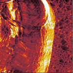
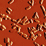
| Phase image of E. coli | Many E. coli | Amplitude E. coli |
 |
 |
 |
|
Silicon grid showing |
Phase image of spores |
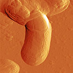
Cell growing from spores - 3D |
 |
 |
|
| Leishmania cell | Epithelial cell |
Protocols
- Startup Procedure
- Shutdown Procedure
- Insertion and laser alignment of probes
- Scanning in contact "non-vibrating" mode
- Approaching in vibrating mode
- Scanning in tapping or "vibrating" mode
- Opening files: We recommend using Gwyddion version 2.22 or later.
- These protocols are currently in development, meanwhile, here is a link to the latest version of the protocols word document for use of "Long Beach": TTAFM_protocols.doc