
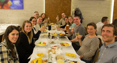
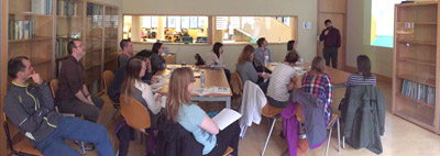
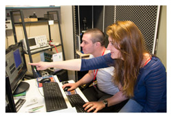
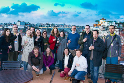
Wednesday was a beautiful sunny day so fortunately all the students had either the morning or afternoon off to make the most of the trip to the beautiful city of Porto. The practical classes went really well, and some of the images obtained by the students can be observed on this page. We always run a “best image” competition, so the students are extra-motivated to collect great AFM data. The results of this competition will be run very shortly. I was particularly impressed that within a classroom environment, with four people around, the students were able to image DNA; getting decent images of structure that are less than one nanometre in height!
- Details
- Hits: 5516
As usual, during our AFM training course , we ran an image competition, challenging the students to submit images they had acquired or processed during the course.
This year, the standard was very high, and we could not decide between two of the entries, so two prizes have been awarded.
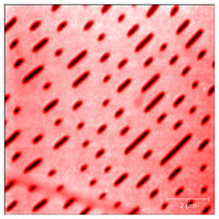
The overall prize goes to the image below of E. coli bacteria which was produced by Dr. Cassandra Terry of UCL, London. The processing on this makes was excellent!
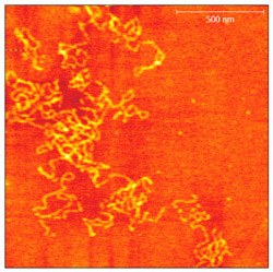
We could not go without giving a prize to this image of double-stranded DNA molecules obtained by Dr. Elin Moe of ITQB, Lisbon. This image, below, won our technical merit prize. Acquiring such an image in your first session with an AFM is impressive!
Congratulations to both, your prizes will be with you soon!
- Details
- Hits: 6553
I am currently writing a chapter for a new book about AFM. The chapter will address AFM issues and artifact. If you have an image that illustrates well an AFM artifact or imaging problem, send it to me, and I might include it in the book!
I will ensure all images are properly credited to their submitters.
Meanwhile if you have an image with a problem, and you cannot identify it, you can also sen that in, and I'll see if I can diagnose the problem.
All images can be sent to This email address is being protected from spambots. You need JavaScript enabled to view it..
- Details
- Hits: 6748
Atomic force microscopy (AFM) is an ideal technique to characterise topography, as well as many other properties, of polymer films and artifacts. AFM has several advantages over electron microscopy in this regards. For instance, most polymers are insulators, and would therefore need to be coated for SEM observation, potentially changing their texture. AFM also does not need a thin sample, as TEM does.
There are many different experiments and modes appropriate for polymer samples. For good reviews see the bibliography at the end of the this article, or the longer article in my book. This article discusses two common types of measurements of polymer samples by AFM.
Phase imaging
Modes such as phase imaging are very useful for polymer composites, and copolymers since they consist of mixed materials, and the distribution of the phases is usually critical for their properties.
As described in section 3.2.3.2 of the book, phase imaging is sensitive to viscoelastic properties of the sample and to tip-sample adhesion. This means that many materials can be differentiated by phase imaging Because of its ability to distinguish many materials, phase imaging has been applied to an enormous number of samples, just some examples include differentiation of semiconductor films, nanoparticle characterisation and counting, observation of spherulites in polymer crystallisation, polymer blend and composite composition, protein adsorption to biomaterials, self assembled monolayers, and many more systems.
Phase imaging is particularly useful for distinguishing features in polymer films which do not exhibit great height contrast. For example, in the image below.
- Details
- Hits: 17835
On this page, I'll link to the occasional articles I write about applications of AFM in different areas
- AFM In Nanotechnology
- Applications of AFM in Nano-science and Nanotechnology
- AFM in Polymer Science
- Applications of AFM in the study of polymer films and plastic products.
- Details
- Hits: 19634
Subcategories
Page 20 of 21