
Site Upgrade
UPDATE: The site has been upgraded to new back-end code. This should help stability and enable new features in the future. Hopefully all the old pages are working now on the new code. If you see any problems, let me know!
The design of the site was changed a little, it looks kind of plain now, maybe I'll revert to the old look later.
There is now a new AFM Image Gallery, which contains more images than before, organised in categories and a few by instrument.
- Details
- Hits: 5700
Free AFM Poster
I have this poster up in the AFM lab here. I made it to save me drawing out how AFMs work everytime I need to explain it to new students or visitors! I saw someone photographing it the other day, so I though I'd make it available here. Click the image below to get the .pdf of the full poster.
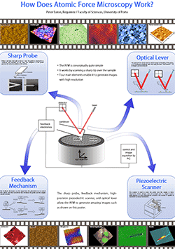
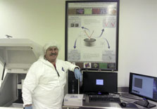
If you print this out and put in your own lab, please send me a photo of the lab! If you do want to print it, you can always This email address is being protected from spambots. You need JavaScript enabled to view it. for a higher-resolution version! The images in this poster are all my own work, and cannot be reproduced except for the purposes described here. UPDATE: Pleased to see the poster in situ in Edward Basgall's microscopy lab at Drexel University in Philadelphia, Thanks, Ed! See below.
- Details
- Hits: 14057
Book Extract - Bacterial Measurements
This article contains a small extract from Chapter 7 of “Atomic Force Microscopy”. Chapter 7 contains descriptions of applications of AFM in materials science, chemistry and physics, biology and the life sciences, nanotechnology, and in industry. This short section describes some examples of applications of AFM in bacteriology. References lists, and the second figure can be found in the full book.
AFM is a highly suitable tool to examine bacteria, and has been widely applied to their study. Bacteria are commonly studied by optical microscopy, which can give an overall idea about gross cell morphology (via a two-dimensional projection), and is also useful for cell-counting studies. In comparison, AFM is slower, and thus is less useful for quantitative cell-counting, but allows measurement of a variety of other cellular properties, particularly by nanoindentation and force spectroscopy experiments [611]. In addition, the greatly increased resolution of AFM allows for the imaging of finer details of cell morphology and sub-cellular features such as pili and fimbriae [612]. The three dimensional information from AFM can also be useful in differentiating morphologies which would look the same in optical microscopy [6]. Various other micro-organisms have been studied by AFM such as spores [178, 613–615], fungi [616, 617], including yeasts [171, 618], viruses [287, 619], and others [620] but here we concentrate on bacteria for the sake of brevity.

Fig. 7.20. Studies of bacterial morphology. Top left: Streptococcus, showing typical linear clusters. Top right: large clusters of Staphylococcus aureus. Bottom left: Salmonella biofilm showing pili-like fimbrial structures. Bottom right: E. coli. All these images were measured in air. Reproduced with permission from [624] (top left) and [626] (bottom left). Right hand images the author's own work.
- Details
- Hits: 9917
Review of our 2014 AFM course

- Details
- Hits: 9524
2014 course is now full
Our 2014 AFM course has had all the places reserved. I am pleased to see we have 16 students from all over the world, including the USA; Malaysia, Germay, Spain, the Czech Republic, Poland, and here in Portugal.
Meanwhile, I will be teaching on another upcoming course, in July 2014, at Kent State University, in Ohio. This course will be 5 days, with full 5 afternoons of instrument time. Places are very limited. More details can be found here: http://afmworkshop.com/atomic-force-microscope-workshop.php. The AFMWorkshop website also hosts a PDF flyer.
- Details
- Hits: 5943
Page 7 of 12