I recently received some praise for my book, and I thought I'd share some reviews with you. Alexander Kraft, from Wacker Chemie AG; said the book represents " A good possibility for beginners in AFM to understand the basics and to gain a deeper insight in this measurement method." Thanks, Alexander! Here are some more quotes:
"A super-clear, easy-to-read, informative, and intuitive introduction to AFM, the best I have found. Normally, I find that books like this can be a bit dense and/or skip over details of how things work, but this book builds everything up intuitively and with such clarity it'd probably be able to be understood by a freshman college student--but, without sacrificing the necessary detail."
- Reviewer at Amazon.com
"Atomic Force Microscopy provides the basic knowledge necessary for successful AFM operation while avoiding the trap of providing more detail than beginners can handle. It boasts seven chapters, each of them accessible and self-contained; readers can thus cherry-pick the topics of relevance for their specific problems. After a short introduction about the historical background and the contemporary context, the book covers practical issues such as understanding AFM design; working in operational modes; measuring, processing, and analyzing AFM images; and spotting and avoiding artifacts. For readers inclined to explore further uses, the book's last chapter discusses various applications that illustrate the multitude of measurement options available with AFMs...Atomic Force Microscopy is a great introduction to AFMs for beginners and, although light on theory, also serves as a good starting point for more serious users."
- Udo D. Schartz, in Physics Today
"I recommend this book to any reader who wants to enter the world of force microscopy. This book is easy to read, entertaining, with a practical approach that allows, after their reading, have a realistic idea and practice of this technique. This book touches on all the points and issues that are critical to understanding the proximity microscopy.
These include instrumentation, measurement modes, familiarization with the images, the routine procedures for image processing, one section devoted to artefacts and finally potential applications of the technique. From my point of view, is one of the books on microscopy of proximity, which is easier to read and with a high applicability in measuring routines."
- Carmen Serra, Nanotechnology and Surface Analysis Service, University of Vigo
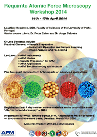
Finally, a reminder: in April 2014, We will be giving another of our successful AFM training course. At the time of writing there is ONE places left on the course. I recommend anyone interested to sign up as soon as possible. Click the image below for more information.
- Details
- Hits: 13578
Our 2014 AFM course has had all the places reserved. I am pleased to see we have 16 students from all over the world, including the USA; Malaysia, Germay, Spain, the Czech Republic, Poland, and here in Portugal.
Meanwhile, I will be teaching on another upcoming course, in July 2014, at Kent State University, in Ohio. This course will be 5 days, with full 5 afternoons of instrument time. Places are very limited. More details can be found here: http://afmworkshop.com/atomic-force-microscope-workshop.php. The AFMWorkshop website also hosts a PDF flyer.
- Details
- Hits: 7623
This article contains a small extract from Chapter 7 of “Atomic Force Microscopy”. Chapter 7 contains descriptions of applications of AFM in materials science, chemistry and physics, biology and the life sciences, nanotechnology, and in industry. This short section describes some examples of applications of AFM in bacteriology. References lists, and the second figure can be found in the full book.
AFM is a highly suitable tool to examine bacteria, and has been widely applied to their study. Bacteria are commonly studied by optical microscopy, which can give an overall idea about gross cell morphology (via a two-dimensional projection), and is also useful for cell-counting studies. In comparison, AFM is slower, and thus is less useful for quantitative cell-counting, but allows measurement of a variety of other cellular properties, particularly by nanoindentation and force spectroscopy experiments [611]. In addition, the greatly increased resolution of AFM allows for the imaging of finer details of cell morphology and sub-cellular features such as pili and fimbriae [612]. The three dimensional information from AFM can also be useful in differentiating morphologies which would look the same in optical microscopy [6]. Various other micro-organisms have been studied by AFM such as spores [178, 613–615], fungi [616, 617], including yeasts [171, 618], viruses [287, 619], and others [620] but here we concentrate on bacteria for the sake of brevity.

Fig. 7.20. Studies of bacterial morphology. Top left: Streptococcus, showing typical linear clusters. Top right: large clusters of Staphylococcus aureus. Bottom left: Salmonella biofilm showing pili-like fimbrial structures. Bottom right: E. coli. All these images were measured in air. Reproduced with permission from [624] (top left) and [626] (bottom left). Right hand images the author's own work.
- Details
- Hits: 17980
Up until now, this website has been funded by Google text adverts that appear on the right of the page. I don’t control what ads appear there, except that I can remove some categories of ads. Beginning soon, I expect to have actual ads from AFM companies appearing there.
The advertisers do not influence any of the other text I write here. Although I do work with some AFM manufacturer’s equipment more than others (and my co-author on the book, Paul West has been owner/CEO of various AFM companies), I do not really favour instruments of one manufacturer over others. I have used many (more than ten) different AFM instruments over the years and this has led me to think that -
ALL AFM instruments can produce great results.
What is necessary to get great results are a certain level of skill on the part of the operator, a good probe, careful sample preparation, patience, use of the right modes and settings, and sometimes, a dash of luck! While newer instruments certainly offer amazing new modes, and in some cases lower noise levels, increased ease of use, or faster scanning, in my experience 99% of AFM could actually be done on just about any instrument. In my teaching, I hope I explain things that are useful to users of all instruments. Furthermore, although I am happy to get new listings, and factual corrections for the “Where to buy instruments” and “Probes” and “Calibration artifacts” pages, I do not accept copy written by the companies for inclusion on those pages. Any inaccuracies, or opinions are mine alone.
- Details
- Hits: 6869

- Details
- Hits: 14342
Subcategories
Page 17 of 21