3. Other Artifacts
3.3 Flying Tip
This artifact shows that the AFM is not properly tracking the surface.
In the example below the effect is obvious, but this is not always the case.
Sometimes it is clearer if you look at some of the other signals, like amplitude or phase or deflection images. In the images shown below, note how all the features have a "tail" to their left.

In the example above, the image of nanoparticles shows "tails" on each particle, to the left. The image was recorded scanning from right to left.Typically, the left to right image would show the tails on the right side of each feature. It is typical of this effect that you will get different images in the two directions.
How to avoid it.
If you see this, your feedback parameters are incorrect.
It is usually easy to fix. You generally should increase integral gain, then the proportional gain, you may need to change the setpoint too, or even scan more slowly. See the answer about adjusting AFM feedback parameters here, in in my book.
- Details
- Hits: 21237
Where to buy: AFM Probes - Distributors
This page lists distributors (as oposed to manufacturers) of AFM and other SPM probes. Currently the list is (very) incomplete.
- Details
- Hits: 21307
The book, "Atomic Force Microscopy" by Peter Eaton and Paul West was published in March 2010 by OUP.
There is some information about it at this site, but it's somewhat out of date. In particular the contents listing is not quite correct.
Here is the correct contents of "Atomic Force Microscopy":
Preface
Chapter 1: Introduction
1.1 Background to AFM
1.2 AFM today
Chapter 2: Instrumental Aspects of AFM
2.1 Basic concepts in AFM instrumentation
2.2 The AFM stage
2.3 AFM electronics
2.4 Acquisition software
2.5 AFM cantilevers and probes
2.6 AFM instrument environment
2.7 Scanning environment
Chapter 3: AFM Modes
3.1 Topographic modes
3.2 Nontopographic modes
3.3 Surface modification
Chapter 4: Measuring AFM Images
4.1 Sample preparation
4.2 Measuring contact mode images
4.3 Measuring intermittent contact mode images
4.4 High-resolution imaging
4.5 Force curves
Chapter 5: Image Processing in AFM
5.1 Processing AFM images
5.2 Displaying AFM images
5.3 Analysing AFM Images
Chapter 6: Image Artifacts in AFM
6.1 Probe artefacts
6.2 Scanner artefacts
6.3 Image processing artefacts
6.4 Vibration noise
6.5 Noise from other sources
6.6 Other artefacts
Chapter 7: Applications of AFM
7.1 AFM applications in materials science
7.2 AFM applications in nanotechnology
7.3 AFM applications in the life sciences
7.4 Industrial AFM applications
Appendix A: AFM Standards
Appendix B: Scanner Calibration and Certification Procedures
Appendix C: Third Party AFM Software
Index
To buy the book, visit Amazon.com
- Details
- Hits: 40418
AFM Artifacts
1.1 Tip-sample convolution
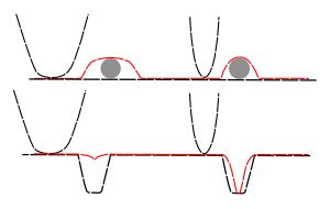
This is an inherent feature of AFM and can never be fully removed. Any AFM image is a convolution of the shape of the probe, and the shape of the sample. This has the effect of making protruding features appear wide, and holes appear smaller (both narrower and often less deep, too). Broader (less sharp) probes will enhance the effect, as shown below.

Illustration of convolution of AFM probe and sample giving rise to the image (red).
Probe-sample convolution tends to have the greatest effect on features of similar or smaller radii than the probe. it can be reduced by deconvolution techniques, discussed further in "Atomic Force Microscopy".
- Details
- Hits: 41203
At the Atomic Force Blog, I publish occasional opinion articles about new things in AFM that catch my eye. Click below to be redirected to the blog.
http://atomicforceblog.blogspot.com/
- Details
- Hits: 32914
Subcategories
Page 10 of 21
